
 by
by Spinal Imaging Techniques: Pushing Boundaries of Detection
Advanced imaging techniques are revolutionizing the field of spinal care. MRI and CT scans provide detailed pictures of the spine and surrounding structures without invasive procedures. These non-invasive modalities allow clinicians to accurately detect and diagnose conditions.
MRI utilizes strong magnetic fields and radio waves to visualize soft tissues with great clarity. It is considered the gold standard for spinal imaging due to its superior soft tissue contrast resolution. MRI easily distinguishes between discs, ligaments, spinal cord and surrounding nerves. It detects herniated discs, spinal stenosis, tumors and infections without exposing patients to radiation.
CT scanning combines X-rays taken from different angles to generate cross-sectional images. While MRI shows soft tissues better, CT clearly depicts bones. It is very effective for imaging fractures, deformities and postoperative changes after spinal fusion surgery. CT scans with contrast dye can help identify tumors and infections.
Image Fusion Merges Data for Enhanced Viewing
Image fusion software superimposes images from different modalities like MRI and CT scans to provide a merged multidimensional view. This integrated visualization allows clinicians to assess bone structures and soft tissues simultaneously on a single image.
Fused images guide precise diagnosis and enable detailed pre-surgical planning. Surgeons can view the relative positions of vertebrae, discs, nerves and underlying vascular structures for planning instrument insertion paths or spinal decompression procedures. Image fusion improves navigation during complex spine surgeries.
Postoperative scans fused with preoperative images facilitate evaluation of surgical outcomes. Clinicians can accurately assess fusion status, hardware placement, decompression extent and residual compression on merged scans. Fusion imaging plays a vital role in evaluating treatment effectiveness and planning further interventions.
Advancement in Software Drives Automated Quantitative Analysis
Latest artificial intelligence and automated image analysis software extract quantitative measurements from scans to enhance diagnostic accuracy. Advanced algorithms identify and segment anatomical structures with high reproducibility.
By automatically measuring disk heights, spinal canal dimensions, nerve root exit angles and vertebral dimensions, these software programs provide objective data to assess degeneration, instability and stenosis severity. They offer automated classification of compression fractures and detection of subtle lesions missed on visual analysis alone.
Integrating demographic data and symptoms, AI can now suggest likely diagnosis based on imaging findings. Automated monitoring of disease progression over time through serial scans will help optimize treatment. As datasets grow, machine learning will continually refine algorithms to aid clinicians.
Emerging Techniques Expand Functional Imaging Capabilities
Novel modalities are enriching functional assessment of the dynamic spine. Diffusion tensor imaging uses MRI to map white matter tracts and nerve pathways in vivo. It assists pre-surgical planning for tumor resections near spinal cords.
Functional MRI detects real time activation patterns during motor and sensory tasks to locate origins of radicular pain. Spinal cord fMRI aids diagnosis of myelopathies by mapping gait, hand function and bladder control centers.
Magnetic resonance neurography provides high resolution imaging of peripheral nerves from their origins in the spinal cord through the paravertebral regions. It diagnoses nerve impingement and entrapment better than conventional MRI.
Positron emission tomography combined with CT quantifies metabolic activity to detect infections, tumors and inflammatory lesions earlier. Single photon emission computed tomography identifies unstable vertebrae at risk of fractures through bone scanning.
These cutting-edge modalities broaden understanding of spinal biomechanics and functional anatomies, fueling advancements in minimally invasive treatments.
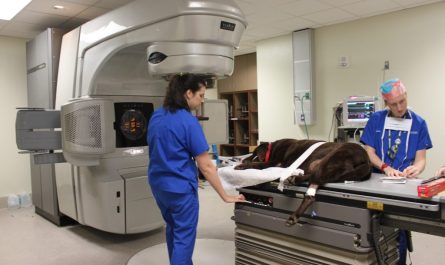
Image-guided Surgery Enhances Precision and Safety
Integration of preoperative scans with intraoperative Spinal Imaging enhances precision and safety of spinal procedures.
Electromagnetic and optical tracking systems allow surgeons to view virtual 3D representations of patient anatomy overlaid on surgical fields. Real-time positional feedback assists accurate instrument insertion and decompression in desired anatomic planes under direct visualization.
Navigation reduces risks from surgical misalignments, over-resections and collateral tissue damage. Combined with robotics, it paves way for minimally invasive spinal surgeries through tubular retractors or small incisions with precision impossible through freehand techniques.
Image guidance increases reproducibility of surgical outcomes. It also enables fusion of postoperative scans with preoperative anatomy models for objective evaluation of treatment outcomes without additional radiation exposure to patients.
With advancements in spinal imaging transforming diagnosis and treatment, integration of quantitative data with artificial intelligence promises even more personalized optimization of care in future. While challenges remain, multidisciplinary research continues fueling new innovations to alleviate the immense worldwide burden of spinal disorders.
Note:
1.Source: Coherent Market Insights, Public sources, Desk research
2.We have leveraged AI tools to mine information and compile it